Female Gamete (Egg) Development
Explore the development and structure of the female gamete, also known as an ovum or egg. This quiz covers the process from a primary oocyte to fertilization.
Pages: 10 Questions per page: 1
Rate this quiz
☆
☆
☆
☆
☆
Avg: 0.00 ( 0 )
Score: 0
Page 1 / 10
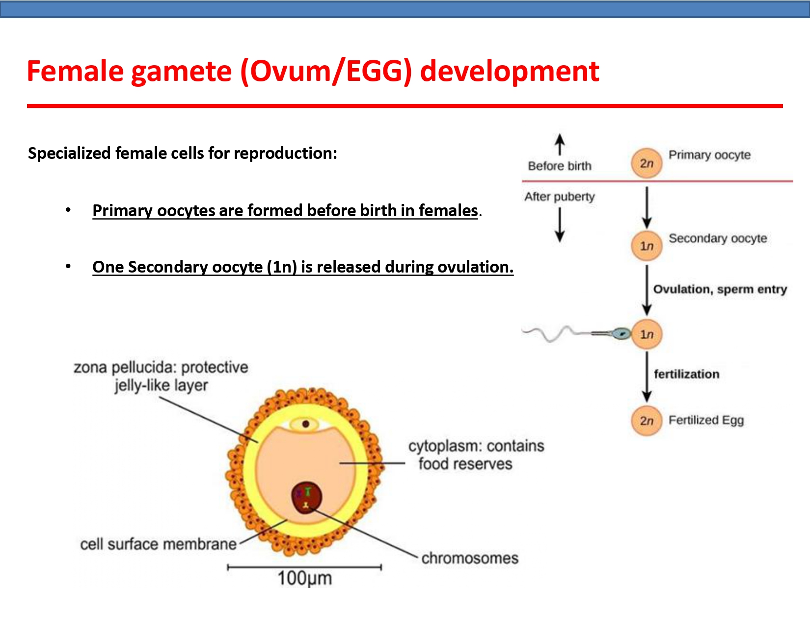
1. According to the provided image, what is the ploidy level of the oocyte released during ovulation, and when are primary oocytes formed?
Explanation: The image states in a bullet point that 'One Secondary oocyte (1n) is released during ovulation,' indicating it is haploid. It also states that 'Primary oocytes are formed before birth in females.' The diagram on the right visually confirms both of these facts.
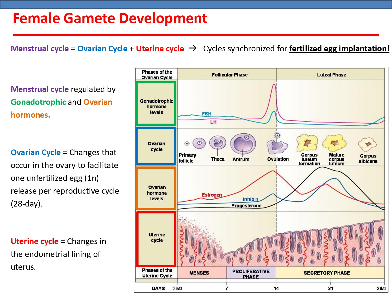
2. According to the diagram, which hormonal event is the most direct trigger for ovulation, which occurs around day 14?
Explanation: The diagram shows a dramatic spike (surge) in LH levels peaking just before day 14, immediately preceding the event labeled 'Ovulation'. This LH surge is the primary trigger for the mature follicle to rupture and release the egg. Progesterone levels are low at this time and rise only after ovulation, while estrogen peaks just before the LH surge, helping to trigger it.
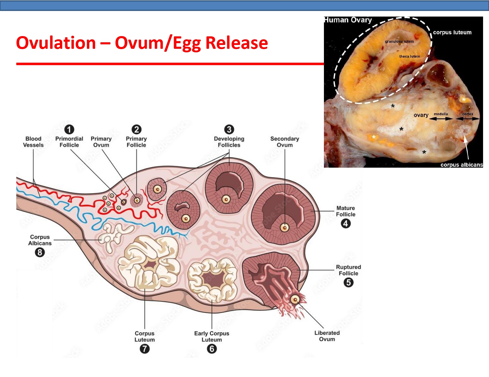
3. Based on the provided diagram and photograph, which numbered structure develops from the ruptured follicle after ovulation and corresponds to the large, yellowish, hormone-producing structure clearly labeled in the photograph?
Explanation: After the mature follicle (4) ruptures and releases the ovum (5), the remaining follicular cells develop into the corpus luteum (6 and 7). The photograph shows a large, yellowish structure identified as the corpus luteum, which is responsible for producing progesterone. The diagram shows the mature corpus luteum at stage 7. The corpus albicans (8) is the degenerated, scar-like remnant of the corpus luteum.
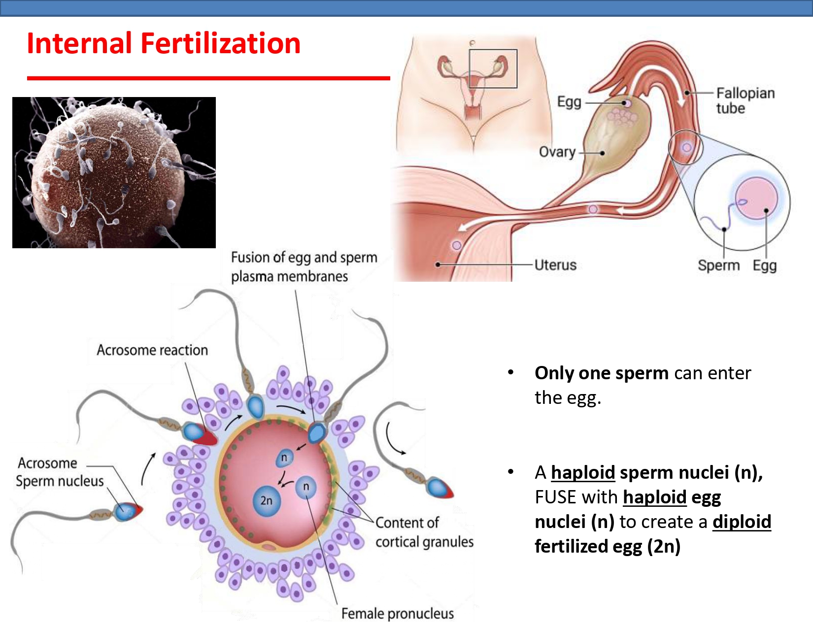
4. According to the diagram, what is the direct result of the fusion of a haploid sperm nucleus (n) with a haploid egg nucleus (n)?
Explanation: The diagram and accompanying text explicitly state that a haploid (n) sperm nucleus fuses with a haploid (n) egg nucleus. This combination of genetic material results in the formation of a single diploid (2n) cell, which is the fertilized egg or zygote.
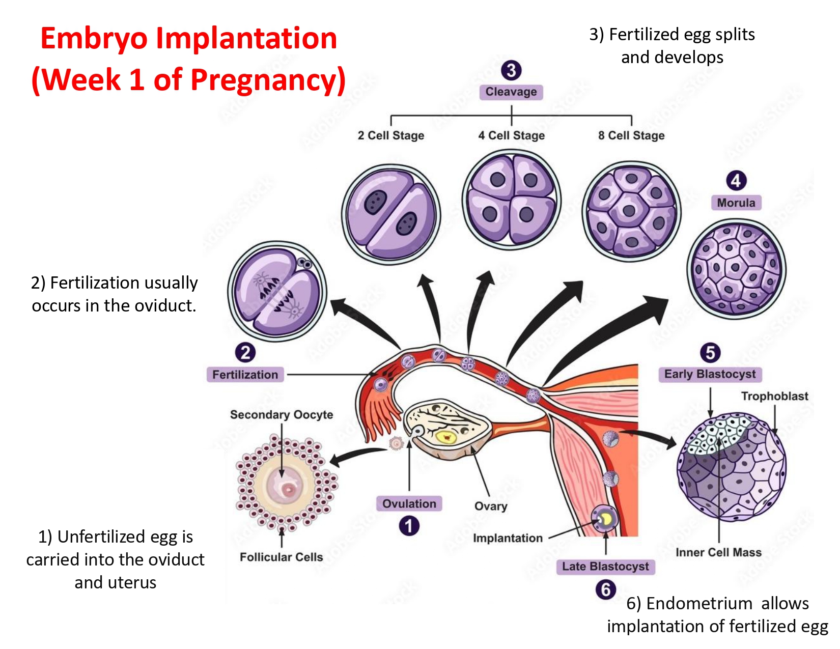
5. According to the diagram, what is the correct sequence of developmental stages for the embryo after fertilization?
Explanation: The diagram shows that after fertilization (step 2), the fertilized egg undergoes cleavage (step 3), where it divides into 2, 4, and 8 cells. This ball of cells then becomes a morula (step 4). Following the morula stage, it develops into a blastocyst (step 5), which is the stage that implants into the uterine wall.
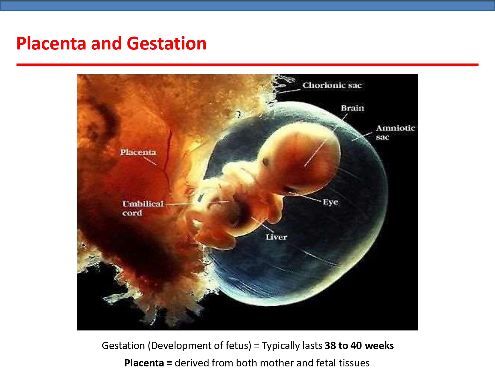
6. According to the information provided in the image, from what is the placenta derived?
Explanation: The text at the bottom of the image explicitly states, 'Placenta = derived from both mother and fetal tissues'.
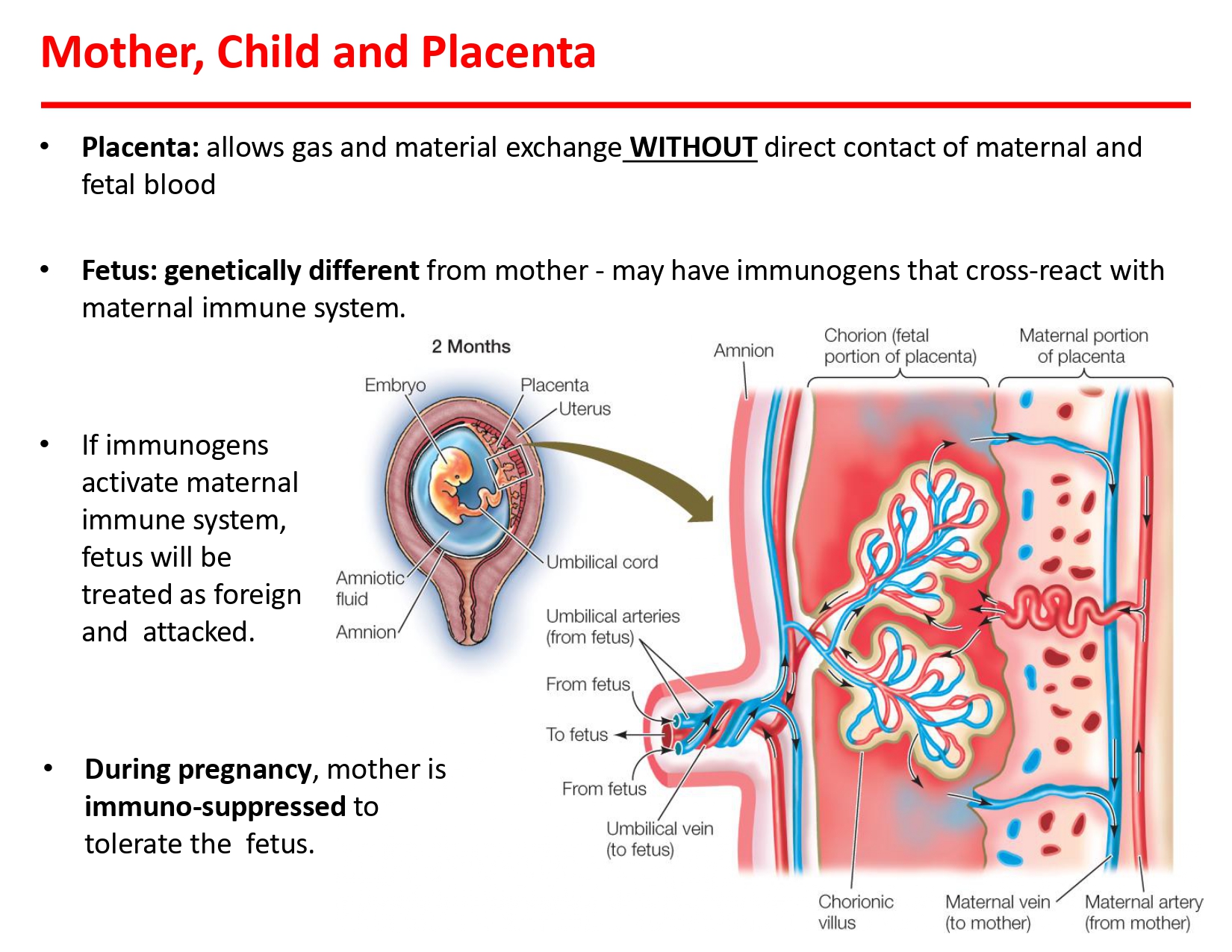
7. Based on the provided information, which statement accurately describes the function of the placenta?
Explanation: The first bullet point on the slide explicitly states that the placenta "allows gas and material exchange WITHOUT direct contact of maternal and fetal blood." The detailed diagram of the placenta confirms this by showing that fetal blood vessels within the chorionic villi are bathed in pools of maternal blood, but the two circulatory systems do not mix.
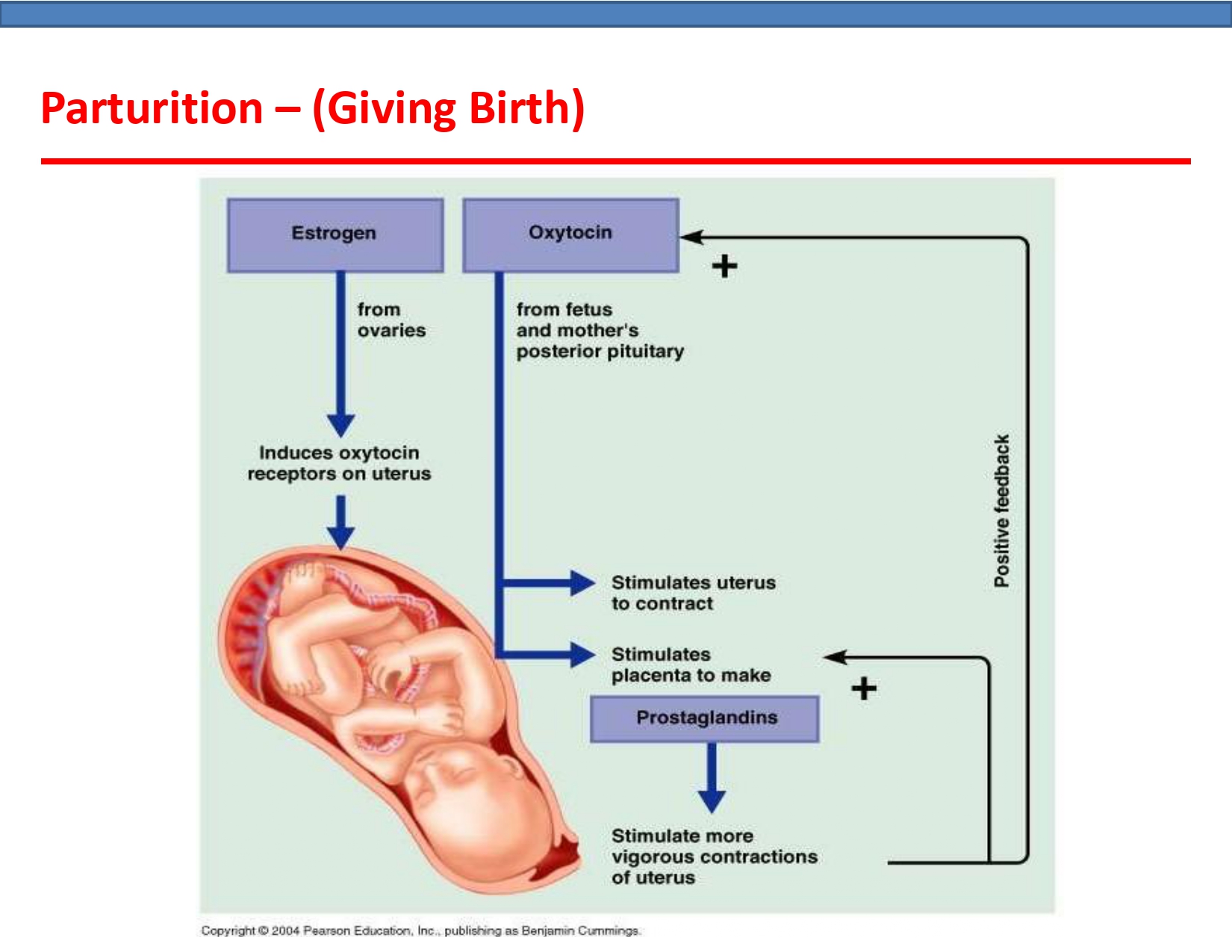
8. According to the diagram, the process of parturition (giving birth) is driven by a positive feedback loop. Which hormone is shown to be central to this feedback mechanism, leading to progressively stronger uterine contractions?
Explanation: The diagram illustrates a positive feedback loop where uterine contractions stimulate the release of more oxytocin. This increased oxytocin then stimulates even stronger contractions, perpetuating the cycle until birth is complete. While prostaglandins also stimulate contractions, oxytocin is depicted as the key hormone in the feedback signal to the mother's posterior pituitary.

9. Based on the provided diagrams, what is the correct chronological order of the fetal head movements during parturition after the onset of labor?
Explanation: The image displays the stages of parturition in sequential order from left to right, top to bottom. After the 'Onset of labour', the first movement shown is 'Flexion'. This is followed by 'Internal rotation of head'. Next, as the head emerges, it undergoes 'Extension'. Finally, after the head is delivered, it performs an 'External rotation of head'.
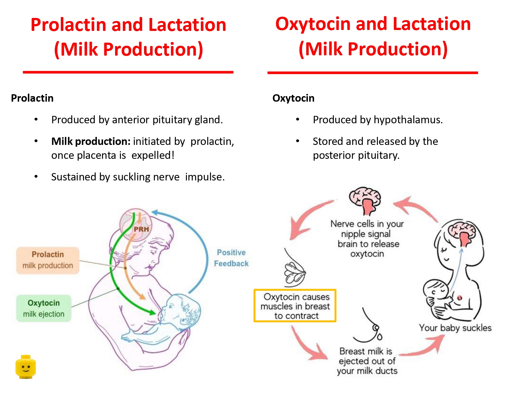
10. Based on the information provided in the image, what is the primary difference between the functions of prolactin and oxytocin in lactation?
Explanation: The image clearly distinguishes the roles of the two hormones. The section on Prolactin states its function is 'Milk production'. The section on Oxytocin describes a process where it 'causes muscles in breast to contract' leading to milk being 'ejected out of your milk ducts'. The summary diagram on the left also labels prolactin's effect as 'milk production' and oxytocin's as 'milk ejection'.